
🧪 Leukemia Diagnosis — Complete Patient Guide
A medically accurate, step-by-step guide to the entire leukemia diagnostic process — from the first blood test to bone marrow biopsy, molecular profiling, and understanding what your results mean.
The Leukemia Diagnostic Journey
A leukemia diagnosis is rarely immediate. It unfolds through a structured sequence of tests, each building on the last, from an initial blood test that raises suspicion to comprehensive molecular profiling that defines the precise leukemia subtype and guides every treatment decision. For most patients, this diagnostic journey takes between two and four weeks — though acute leukemias require urgent evaluation and treatment may begin within days of initial blood test abnormalities.
Understanding each step of this process helps patients participate more confidently in their own care, interpret results meaningfully, and ask the right questions of their medical team. Receiving a leukemia diagnosis is one of the most significant events in a person's life; knowing what each test means and why it is needed transforms a frightening, opaque experience into a comprehensible medical process with clear next steps.
The diagnostic evaluation not only confirms the presence of leukemia but also defines its precise molecular identity — information that is inseparable from treatment selection. Unlike many cancers where the diagnosis and the treatment plan are sequential, in leukemia they are partially simultaneous: certain molecular results (such as APL or BCR-ABL1 positivity) can change the treatment initiated within hours of their return.
Step-by-Step Diagnostic Pathway
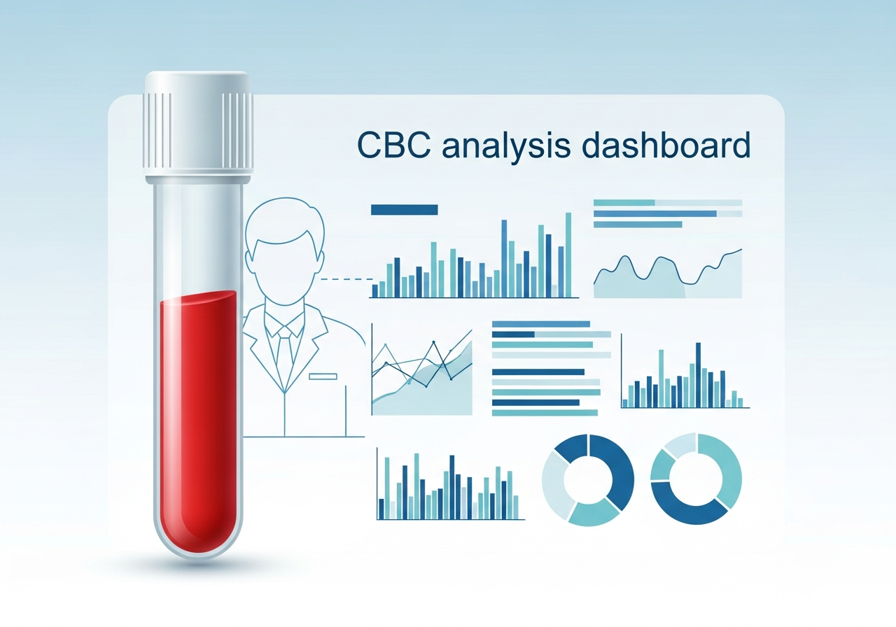
- Complete Blood Count (CBC) with differential: The first and most important screening test. Measures white blood cells (WBC), red blood cells (RBC), hemoglobin, hematocrit, and platelets. A differential counts the types of white cells. Abnormal results — including very high or very low WBC, anemia, or low platelets — prompt further investigation. Read our complete guide to blood tests in leukemia diagnosis.
- Peripheral blood smear: A drop of blood examined under microscope by a pathologist. Can reveal leukemia blasts (immature cells) directly in the bloodstream, which are diagnostic of acute leukemia in many cases. Also reveals cell morphology abnormalities (pelger-huet anomaly, hypersegmentation) suggesting bone marrow disease.
- Hematologist-oncologist referral: Any patient with suspected leukemia should be seen by a hematologist-oncologist without delay. Acute leukemias require same-day or next-day evaluation. Chronic leukemias should be seen within days to one week.
- Bone marrow biopsy and aspirate: The definitive diagnostic procedure. A needle is inserted into the posterior iliac crest (hip bone) under local anesthesia to collect both bone marrow fluid (aspirate) and a core of bone marrow tissue (biopsy). Results define blast percentage, cell morphology, and provide tissue for all molecular testing. See our complete bone marrow biopsy guide.
- Cytogenetic analysis (karyotype): The leukemia cells are cultured and their chromosomes examined under microscope. Detects large chromosomal changes — deletions, duplications, translocations — that define risk category. The Philadelphia chromosome (t(9;22)) and many others are detected this way.
- Fluorescence in situ hybridization (FISH): More sensitive than standard cytogenetics. Uses fluorescent DNA probes to detect specific gene fusions or deletions. Particularly important for detecting BCR-ABL1, del(17p) in CLL, and other targeted abnormalities.
- Flow cytometry: Analyzes thousands of individual cells for specific surface proteins (CD markers) using laser-based technology. Essential for distinguishing myeloid from lymphoid leukemia, identifying the exact stage of maturation arrest, and diagnosing specific subtypes like CLL, hairy cell leukemia, and mature T-cell leukemias.
- Next-generation sequencing (NGS): Comprehensive gene mutation panel that identifies point mutations in genes such as FLT3, NPM1, IDH1/2, RUNX1, ASXL1, TP53, and many others. These results determine targeted therapy eligibility and contribute to risk stratification.
- Imaging studies: CT scans, PET-CT, and occasionally MRI identify organ involvement, lymph node enlargement, spleen size, and any CNS involvement requiring additional treatment planning.
CBC Findings by Leukemia Type
The pattern of CBC abnormalities provides important early clues about which type of leukemia may be present, even before biopsy results are available:
| Leukemia Type | WBC | Hemoglobin | Platelets | Key Smear Finding |
|---|---|---|---|---|
| ALL (acute) | Very high (often >50,000) or low | Low (anemia) | Low (thrombocytopenia) | Lymphoblasts with prominent nucleoli |
| AML (acute) | Variable; often high with blasts | Low | Low | Myeloblasts; Auer rods (pathognomonic) |
| CLL (chronic) | High (>10,000 lymphocytes) — often incidental | Normal or mildly low | Normal or mildly low | Smudge cells; mature small lymphocytes |
| CML (chronic phase) | Very high (>100,000); full granulocyte spectrum | Normal to mildly low | Often elevated initially | Full myeloid maturation spectrum; basophilia |
Diagnosis Article Library
Blood Tests and CBC in Leukemia Diagnosis
How complete blood counts, peripheral blood smears, and routine labs first detect leukemia — and what abnormal results mean for next steps.
Read more →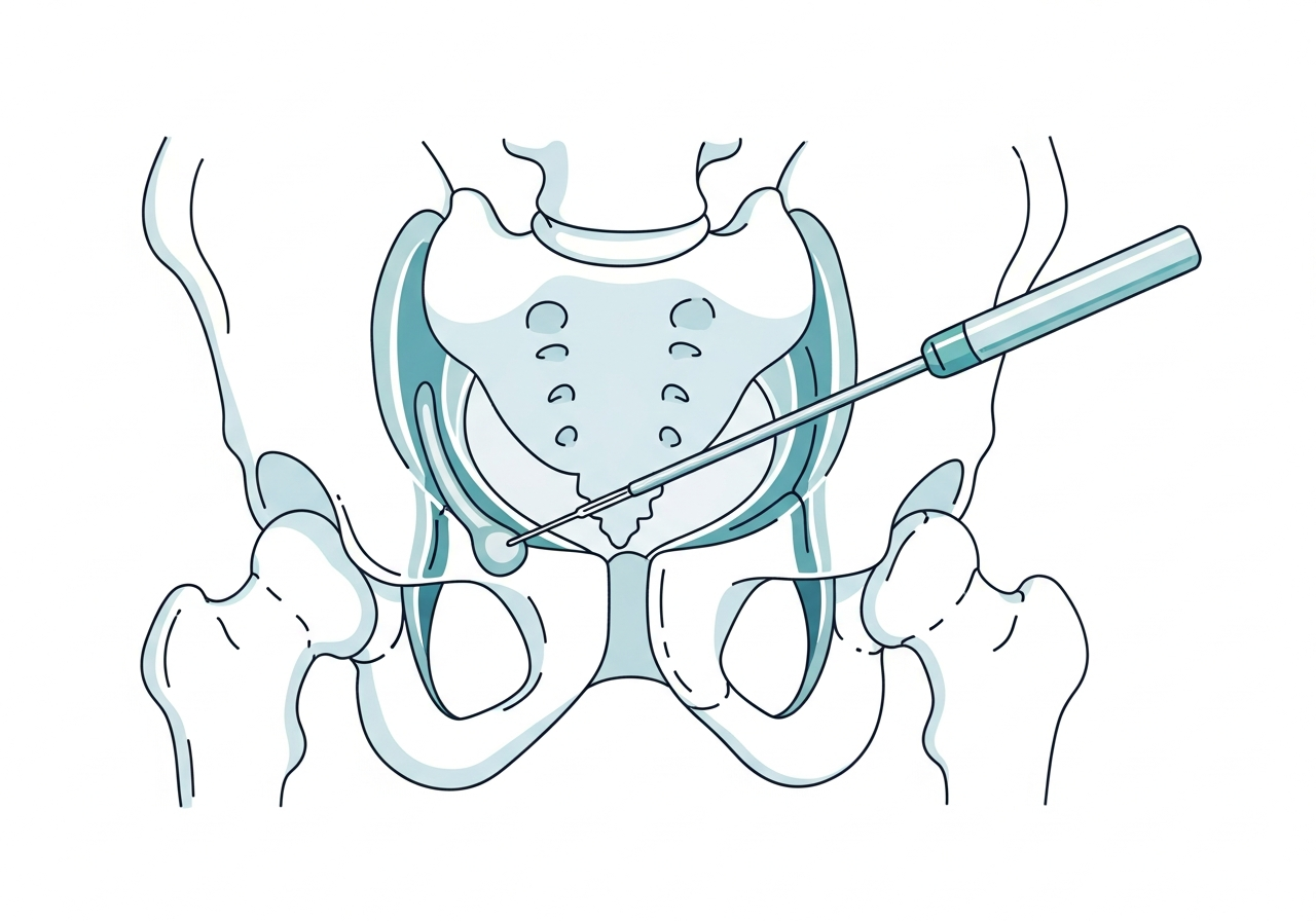
Bone Marrow Biopsy: What to Expect
What happens during a bone marrow aspiration and biopsy, how to prepare, what is tested in the sample, and how results guide treatment.
Read more →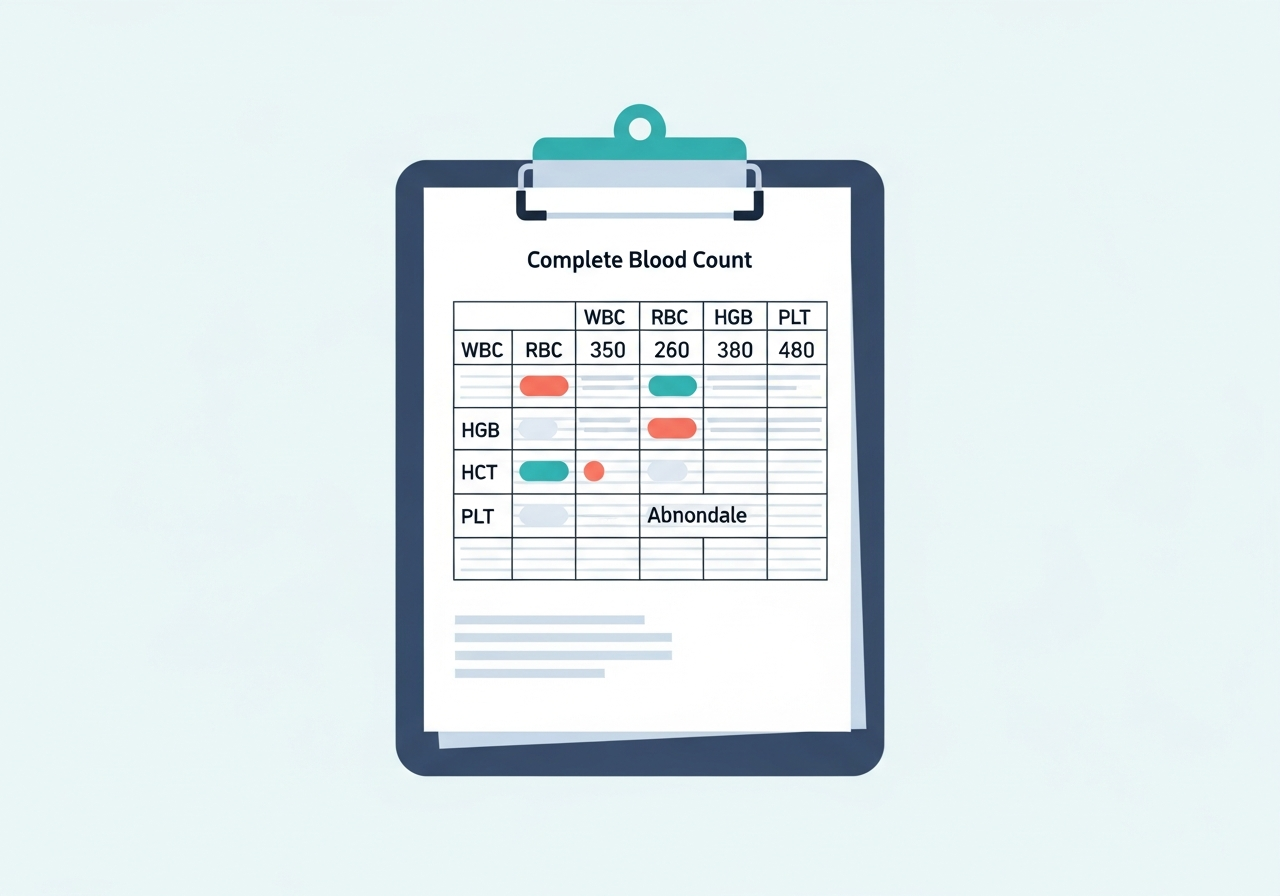
Understanding Your Leukemia Test Results
A patient's guide to decoding CBC values, flow cytometry reports, cytogenetics, and molecular panels after a leukemia diagnosis.
Read more →Featured Guides
Blood Tests for Leukemia Diagnosis
A comprehensive guide to every blood test used in leukemia evaluation — CBC, LDH, uric acid, coagulation studies — what each measures and what abnormal results indicate.
Read full guide →Bone Marrow Biopsy in Leukemia
Everything patients need to know before, during, and after a bone marrow biopsy — the procedure, what information it provides, and how results shape treatment planning.
Read full guide →Understanding Your Leukemia Test Results
Receiving a stack of laboratory reports can be overwhelming. Our guide to understanding leukemia test results walks through each common result type — blast percentage, chromosome findings, mutation reports — and explains in plain language what each means for diagnosis and treatment. Key concepts include: what "complete remission" means and how it is defined; how minimal residual disease (MRD) negativity affects long-term outcomes; and how risk stratification categories (favorable, intermediate, adverse) translate into treatment intensity recommendations. Patients who understand their results engage more effectively with their treatment team and make better-informed decisions at every stage of care.
Leukemia Diagnosis — Frequently Asked Questions
Leukemia diagnosis begins with a Complete Blood Count (CBC) with differential, which screens for abnormal cell counts. Abnormal results prompt a peripheral blood smear (blood under microscope), then a bone marrow biopsy and aspirate. Biopsy tissue undergoes cytogenetic analysis, FISH testing, flow cytometry (cell surface marker analysis), and next-generation sequencing (gene mutation panel) to fully define the leukemia type and molecular profile used for treatment planning.
A CBC result is typically available within hours. Peripheral blood smear review is same-day. A bone marrow biopsy provides initial morphology results within 24–48 hours, which is sufficient to begin treatment in acute leukemia. Full molecular testing (cytogenetics, FISH, NGS) typically takes 10–21 days. Patients with acute leukemia often begin treatment while awaiting complete molecular results.
In most cases, yes. A bone marrow biopsy is the definitive diagnostic test. It confirms the blast percentage (percentage of immature cancer cells), identifies the precise cell type (myeloid vs. lymphoid), provides tissue for cytogenetic and molecular testing, and establishes baseline disease burden for monitoring treatment response. Without it, precise subtype classification and optimal treatment selection are not possible.
An elevated white blood cell count (leukocytosis) is one of the most common CBC findings in leukemia, but it is not specific — it also occurs in infections, inflammatory conditions, steroid use, and physiological stress. What makes leukemia more likely is the pattern and type of white cells elevated, the presence of immature blasts on smear, and accompanying anemia and thrombocytopenia. A physician must interpret the result in full clinical context.
MRD (minimal residual disease) testing is a highly sensitive technique that detects extremely small numbers of residual leukemia cells — as few as 1 in 100,000 or 1 in 1,000,000 normal cells — using flow cytometry or PCR after treatment. MRD negativity (no detectable residual leukemia) is a strong predictor of durable remission and is increasingly used to guide decisions about transplant timing, maintenance therapy, and treatment de-escalation.
Explore Related Topics
This content is for informational and educational purposes only. It is not a substitute for professional medical advice, diagnosis, or treatment. Always consult your physician or a qualified healthcare provider with any questions about a medical condition. Read our full disclaimer.