
📋 Quick Summary
- Topic: Risk factors, aggressive disease course, treatment with intensive chemotherapy and stem cell transplant, and survival data.
- Key Takeaway: Early recognition and medical consultation are critical for the best clinical outcomes.
- Remember: Always consult a qualified healthcare professional if you have concerns.
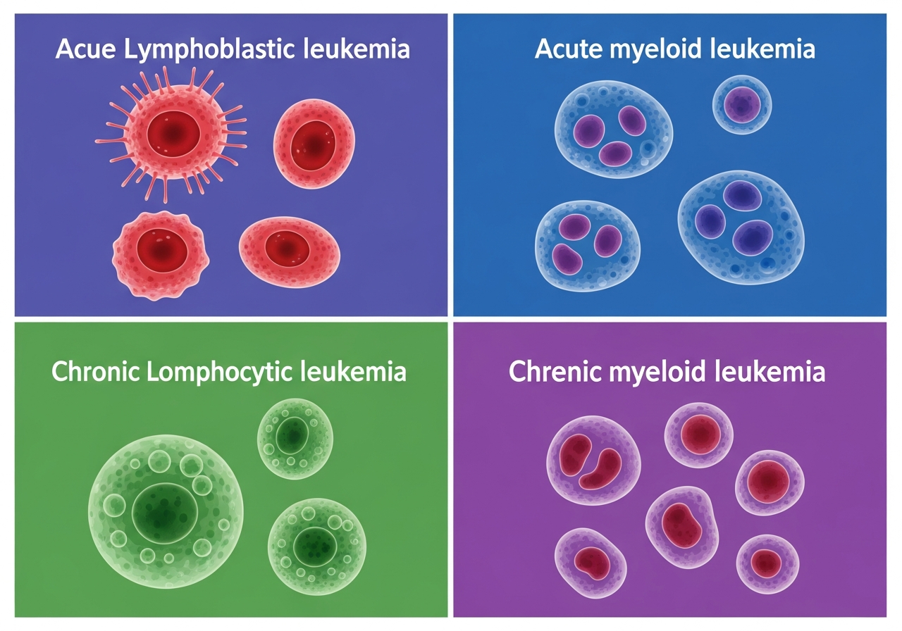
Acute Myeloid Leukemia (AML): A Comprehensive Guide
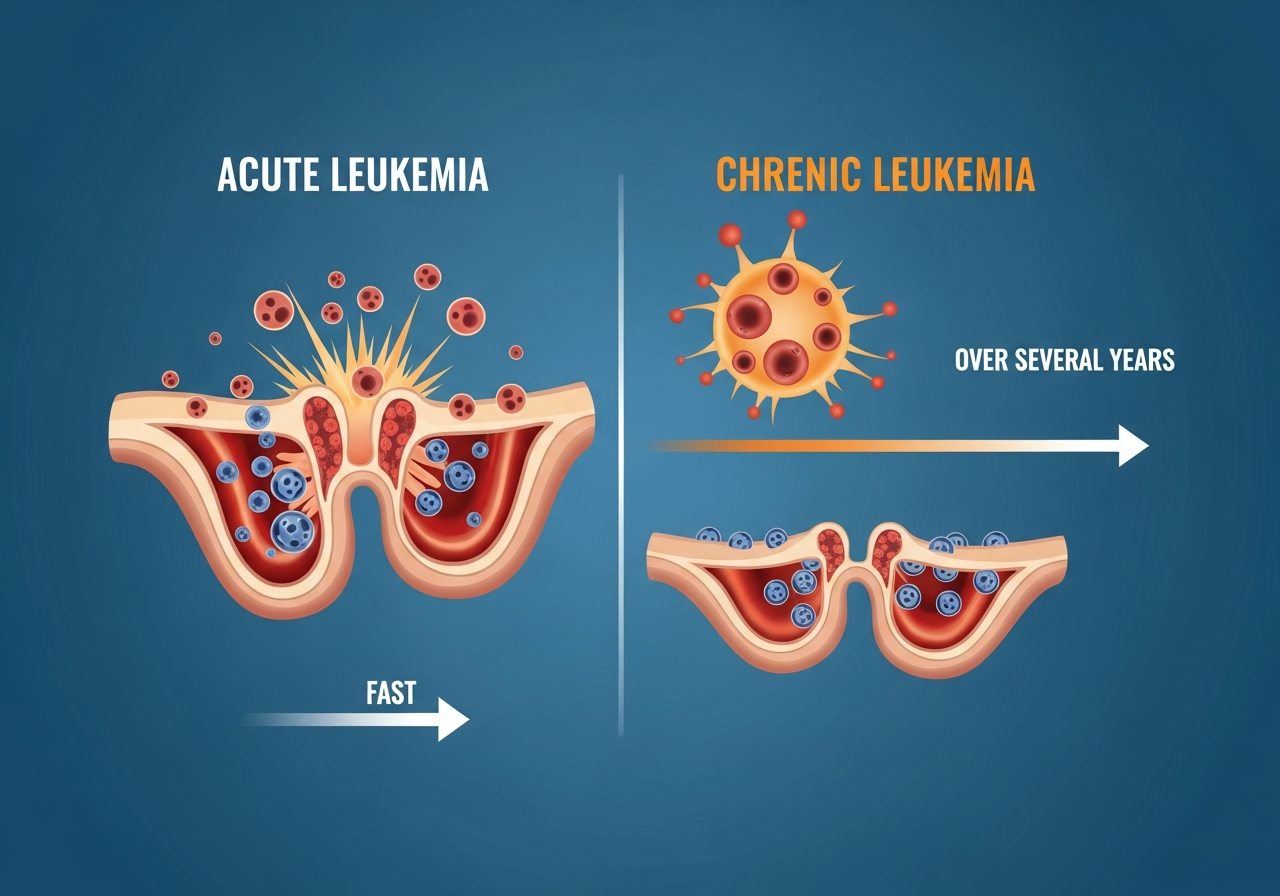
Acute Myeloid Leukemia (AML) is the most common acute leukemia in adults and one of the most challenging cancers to treat. It arises from the malignant transformation of myeloid progenitor cells — the precursors of red blood cells, granulocytes, monocytes, and platelets — and progresses rapidly. Without treatment, AML is fatal within weeks to months. Even with treatment, outcomes remain variable, particularly in older adults and those with high-risk molecular features.
AML is a heterogeneous disease: it encompasses a large number of biologically distinct subtypes, each with different molecular drivers, treatment sensitivities, and prognoses. The revolution in molecular understanding of AML over the past decade has produced an unprecedented wave of newly approved targeted therapies — IDH inhibitors, FLT3 inhibitors, BCL-2 inhibitors, hedgehog pathway inhibitors — that are transforming outcomes for specific patient subgroups.
Approximately 20,000 Americans are diagnosed with AML each year. The median age at diagnosis is approximately 68, and older adults constitute the majority of patients. This demographic characteristic — a disease primarily of older individuals — significantly constrains treatment options, as intensive chemotherapy carries unacceptable toxicity for many patients over 75 with comorbidities.
The Biology of AML
AML originates in a hematopoietic stem cell or early myeloid progenitor that acquires a series of genetic mutations — typically two or more cooperating events — that simultaneously block differentiation and confer enhanced self-renewal and proliferative advantage. The resulting leukemia cells (myeloid blasts) accumulate in the bone marrow, displacing normal hematopoiesis and eventually entering the peripheral blood.
Key biological features of AML include:
- Differentiation block: AML blasts are arrested at various stages of myeloid maturation. In most subtypes, they are immature myeloblasts; in APL (AML-M3), they are promyelocytes; in monocytic AML (FAB M4/M5), they are monocytic precursors.
- Rapid doubling time: AML blasts proliferate quickly, explaining the rapid pace of clinical deterioration without treatment.
- Leukostasis risk: At very high blast counts (>100,000/μL), blasts can obstruct blood vessels in the lungs and brain, causing respiratory failure and neurological emergencies.
- Bone marrow failure: Suppression of normal erythropoiesis, thrombopoiesis, and granulopoiesis produces the characteristic triad of anemia, thrombocytopenia, and neutropenia.
AML Subtypes: FAB and WHO Classification
AML has been classified using two systems:
FAB (French-American-British) Classification
The older FAB system classifies AML based on the morphological appearance of blasts (M0–M7):
- M0: Minimally differentiated AML
- M1: AML without maturation
- M2: AML with maturation
- M3: Acute Promyelocytic Leukemia (APL)
- M4: Acute Myelomonocytic Leukemia
- M5: Acute Monocytic Leukemia
- M6: Acute Erythroid Leukemia
- M7: Acute Megakaryoblastic Leukemia
WHO Molecular Classification (Current Standard)
The modern WHO classification emphasizes genetic and molecular features over morphology, defining AML subtypes by:
- AML with recurrent genetic abnormalities: t(8;21)/RUNX1-RUNX1T1 (favorable), inv(16)/t(16;16)/CBFB-MYH11 (favorable), PML-RARA (APL, highly treatable), BCR-ABL1, RUNX1 mutation, CEBPA biallelic mutation, NPM1 mutation (generally favorable), FLT3-ITD (intermediate-to-high risk)
- AML with myelodysplasia-related changes: Often in older adults with prior MDS; complex karyotype; poor prognosis
- Therapy-related AML (t-AML): Arising after prior cytotoxic therapy or radiation; poor prognosis
- AML, NOS: Remaining cases not fitting other categories
Molecular Risk Stratification
The ELN (European LeukemiaNet) 2022 risk classification divides AML into three groups based on molecular features:
- Favorable: RUNX1-RUNX1T1 fusion; CBFB-MYH11; NPM1 mutated without FLT3-ITD; CEBPA biallelic — 5-year survival 60–70%+ with standard treatment
- Intermediate: NPM1 mutated with FLT3-ITD; RUNX1 mutated; others not in favorable/adverse — variable prognosis
- Adverse: TP53 mutation; RUNX1-RUNX1T1 with KIT mutation; complex karyotype; monosomal karyotype; IDH2-mutated with adverse co-mutations — poor prognosis; aggressive upfront approach including SCT
Risk Factors for AML
Most AML cases arise in people without identifiable predisposing factors. However, several risk factors are recognized:
- Age: Risk increases dramatically with age; median diagnosis age ~68
- Prior myelodysplastic syndrome (MDS) or myeloproliferative neoplasm (MPN): These "pre-leukemic" conditions can transform to AML — called secondary AML
- Prior chemotherapy or radiation (therapy-related AML): Alkylating agents (7–10 years after exposure) and topoisomerase II inhibitors (2–3 years after exposure) both increase AML risk
- Benzene exposure: Occupational exposure to benzene (a component of petroleum products) is a recognized risk factor
- Genetic syndromes: Down syndrome (trisomy 21), Fanconi anemia, Bloom syndrome, Diamond-Blackfan anemia
- Cigarette smoking: A modest but real increase in AML risk
For a full discussion of how risk varies across life stages, see leukemia risk across life stages.
Clinical Presentation of AML
AML presents acutely, with symptoms typically developing over days to a few weeks. The constellation of findings directly reflects bone marrow failure:
- Anemia-related: Profound fatigue, pallor, breathlessness on exertion
- Thrombocytopenia-related: Easy bruising, petechiae, nosebleeds, gum bleeding, prolonged bleeding
- Neutropenia-related: Fever, rapid-onset infections
- Leukostasis (very high WBC): Shortness of breath, confusion, headache, visual disturbances — emergency
- Organ infiltration: Gingival hypertrophy (gum swelling — characteristic of monocytic AML); hepatosplenomegaly; leukemia cutis (skin nodules); CNS involvement (headache, cranial nerve palsies)
- APL-specific: Life-threatening DIC causing simultaneous bleeding and clotting
Diagnosing AML
AML diagnosis requires ≥20% myeloid blasts in the bone marrow (or peripheral blood). The full diagnostic workup:
- CBC: Usually shows leukocytosis (elevated WBC with blasts), anemia, and thrombocytopenia. Some patients present with low WBC (leukopenia). Learn about CBC in leukemia.
- Peripheral blood smear: Auer rods (needle-like crystalline cytoplasmic inclusions) in myeloblasts are pathognomonic for AML and, when abundant (faggot cells), strongly suggest APL.
- Bone marrow biopsy: Essential — confirms diagnosis, provides specimen for flow cytometry, cytogenetics, and molecular testing. What to expect from bone marrow biopsy.
- Cytogenetics (karyotype/FISH): Identifies favorable fusion genes (CBFB-MYH11, RUNX1-RUNX1T1, PML-RARA) and adverse abnormalities (monosomy 5, 7; complex karyotype)
- Molecular panel (NGS): FLT3-ITD and TKD, NPM1, IDH1, IDH2, RUNX1, TP53, ASXL1, CEBPA — required for ELN risk stratification and to identify targets for targeted therapy
- Coagulation studies: PT, PTT, fibrinogen, D-dimer — especially important if APL is suspected
Full interpretation guidance: understanding leukemia test results.
Treatment of AML
Induction Chemotherapy
Standard induction for fit patients is "7+3": continuous infusion cytarabine for 7 days plus an anthracycline (daunorubicin or idarubicin) for 3 days. This achieves complete remission (CR) in approximately 60–80% of younger adults. Older adults may receive lower-intensity alternatives if standard intensity is not tolerated.
Molecularly targeted additions to induction:
- FLT3-mutated AML: Midostaurin added to standard 7+3 (FDA-approved) significantly improves event-free and overall survival
- Gemtuzumab ozogamicin (GO): Anti-CD33 antibody-drug conjugate added to induction for favorable and intermediate-risk AML
- Older/unfit patients: Azacitidine + venetoclax (BCL-2 inhibitor) is the new standard for patients ineligible for intensive chemotherapy
Consolidation Therapy
Patients achieving CR receive consolidation to eliminate residual disease. Options include:
- High-dose cytarabine (HiDAC) — 3–4 cycles, preferred for favorable-risk AML
- Allogeneic stem cell transplantation (allo-SCT) — recommended in first CR for intermediate and adverse-risk patients with a suitable donor
- Targeted agents (IDH inhibitors: enasidenib for IDH2, ivosidenib for IDH1) may be incorporated in maintenance
Targeted Agents in Relapsed/Refractory AML
- Gilteritinib: Second-generation FLT3 inhibitor; FDA-approved for relapsed/refractory FLT3-mutated AML
- Enasidenib (IDH2) / Ivosidenib (IDH1): Well-tolerated oral agents with single-agent activity; increasingly used in combination with azacitidine or chemotherapy
- Venetoclax combinations: Highly active in previously untreated older patients; showing activity in relapsed setting
- Glasdegib (hedgehog inhibitor): Approved in combination with low-dose cytarabine for unfit patients
- Gemtuzumab ozogamicin (GO): Monotherapy or combined with chemotherapy for CD33+ AML
APL: A Unique and Highly Treatable AML Subtype
Acute Promyelocytic Leukemia (APL) deserves special attention because it is simultaneously the most dangerous and the most treatable subtype of AML. APL is defined by the PML-RARA fusion gene, which blocks promyelocyte differentiation and produces a devastating clotting disorder (DIC) while simultaneously conferring exquisite sensitivity to All-Trans Retinoic Acid (ATRA).
ATRA combined with arsenic trioxide (ATO) — without traditional cytotoxic chemotherapy in standard-risk APL — achieves complete remission in over 95% of patients and cure in approximately 90%. This regimen represents one of the most successful targeted therapy combinations in all of oncology. Critically, ATRA should be started immediately upon clinical suspicion of APL — before cytogenetic confirmation — to prevent fatal hemorrhage from DIC.
The prompt recognition of APL through clinical features (bleeding, high-blast AML with possible Auer rods / faggot cells, DIC labs) is therefore a true medical emergency with life-or-death implications for the timing of ATRA initiation.
Prognosis in AML
AML prognosis varies widely by molecular risk category and patient age:
| Risk Category | 5-Year OS (Younger Adults) | Common Approach |
|---|---|---|
| Favorable (APL) | ~90% | ATRA + ATO (no SCT needed) |
| Favorable (non-APL) | 60–70% | HiDAC consolidation (no SCT) |
| Intermediate | 40–55% | Allo-SCT in CR1 if donor available |
| Adverse | 10–25% | Allo-SCT; clinical trial; novel agents |
| Older adults (>70) | 10–20% | Aza + venetoclax; supportive care |
Patient Considerations in AML
AML treatment, particularly induction chemotherapy, requires prolonged hospitalization (typically 4–6 weeks for a single induction course). Patients should prepare for this: arranging family support, childcare or dependent care, and financial/insurance management. Clinical trial enrollment is strongly encouraged — particularly for patients with adverse molecular profiles — and should be discussed immediately at time of diagnosis. See our guide to clinical trials and treatment decisions. Fertility preservation (sperm banking, egg/embryo freezing) must be discussed and arranged BEFORE starting induction chemotherapy.
Caregiver Guidance
AML treatment often begins as a medical emergency — patients may be hospitalized urgently within days of diagnosis. Caregivers should be prepared for rapid decision-making around treatment choices, particularly regarding: intensive vs. non-intensive induction, clinical trial participation, and allogeneic SCT planning. During hospitalization, caregivers provide essential emotional support and serve as advocates within the hospital system. After discharge, the primary caregiver responsibilities shift to infection monitoring (immediately seeking emergency care for any fever), medication management, nutritional support, and transportation to frequent outpatient visits.
When to Seek Emergency Care in AML
- Any newly diagnosed or suspected AML — this is an oncologic emergency requiring immediate hospital-based management
- Fever above 101°F (38.3°C) in any AML patient — febrile neutropenia is life-threatening
- Sudden severe headache, confusion, or neurological symptoms — possible intracranial hemorrhage or CNS involvement
- Bleeding from multiple sites simultaneously — possible DIC (especially in APL)
- Rapidly worsening shortness of breath with very high WBC count — possible leukostasis emergency
🚨 AML Emergencies — Call 911 or Go to Emergency Room For:
- Suspected APL with any bleeding — ATRA must be started immediately
- Fever above 101°F (38.3°C) — febrile neutropenia
- Sudden severe headache or confusion — possible intracranial hemorrhage
- Extreme breathlessness with very high white blood cell count — leukostasis
💬 Questions to Ask Your AML Treatment Team
- What is my ELN risk category based on my molecular testing?
- Do I have FLT3, IDH1/2, or NPM1 mutations, and how do these affect my treatment?
- Am I a candidate for intensive chemotherapy or should I have a less intensive regimen?
- Is an allogeneic stem cell transplant planned, and what is the timeline?
- Are there clinical trials available for my specific AML subtype?
- How should my family be tested for HLA matching for potential stem cell donation?
This content is for informational and educational purposes only. It is not a substitute for professional medical advice, diagnosis, or treatment. Always consult your physician or a qualified healthcare provider with any questions about a medical condition. Never disregard professional medical advice or delay seeking it because of information you have read on this website. Read our full disclaimer.
Frequently Asked Questions
AML progresses rapidly and suppresses production of all normal blood cells, making patients extremely vulnerable to fatal infections and bleeding.
Age (over 65), prior chemotherapy or radiation, genetic disorders like Down syndrome, and benzene or pesticide exposure.
Standard treatment involves intensive induction chemotherapy, consolidation therapy, and often an allogeneic stem cell transplant for eligible patients.